From routine checkups to treatments for surgery, Denton Foot and Ankle Surgery is equipped to handle all your podiatric needs.
FAX : 833-905-2567
c.chappell@dentonfootandanklesurgery.com

Podiatric Services

Foot and Ankle Solutions

When you have an injury or issue with your feet or your ankles, let Denton Foot and Ankle Surgery be your solution! While your general practitioner may be able to offer some assistance, our doctors are highly specialized and fully committed to nothing but your feet and ankles. We offer both general podiatry and surgical options in order to help you heal quickly. From working with you to help strengthen your muscles or treat fungus issues, to cutting-edge surgical solutions, we have the right treatment for your foot and ankle needs.

Select Your Service

Select the issue you are experiencing from our list below to learn more about how we can help with solving your foot and/or ankle issue

Contact Us

Contact us through our website, via email, or call us to set your appointment

Visit Our Clinic

After filling out your new patient paperwork, our podiatrists will review your issues and discuss a treatment plan

Achilles Tendon Rupture

An Achilles tendon rupture is a complete or partial tear that occurs when the tendon is stretched beyond its capacity. Achilles tendon ruptures are most often seen in “weekend warriors” typically, middle-aged people participating in sports in their spare time. Less commonly, illness or medications, such as steroids or certain antibiotics may weaken the tendon and contribute to ruptures. Patients with chronic un-treated Achilles tendonitis can also lead to a weaken tendon and lead to a rupture.

What are the symptoms of Achilles Tendon Rupture?

A person with a ruptured Achilles tendon may experience one or more of the following

These symptoms require prompt medical attention to prevent further damage. Until the patient can see a doctor, the “R.I.C.E.” method should be used

Diagnosis

In diagnosing an Achilles tendon rupture, the foot and ankle surgeon will ask questions about how and when the injury occurred. The surgeon will examine the foot and ankle, felling for a defect in the tendon that suggests a tear. Range of motion and muscle strength will be evaluated and compared to the uninjured foot and ankle. If the Achilles tendon is ruptured the patient will have less strength in pushing down and will have difficulty rising onto the toes. The diagnosis of an Achilles tendon rupture is typically straightforward and can be made through this type of examination. In some cases, however the surgeon may order an MRI or other advanced imaging tests.

Treatment

Treatment options for an Achilles tendon rupture include surgical and non-surgical approaches. The decision of whether to proceed with surgery or non-surgical treatment is based on the severity of the rupture and the patient’s health status and activity level.

Non-Surgical Treatment

Is generally associated with a higher rate of re-rupture is selected for minor ruptures, less active patients and those with medical conditions that prevent them from undergoing surgery. Non-Surgical treatment involves the use of a cast, walking boot and brace to restrict motion and allow the torn tendon to heal.

Surgical Treatment

Surgery offers important potential benefits. Besides decreasing the likelihood of re-rupturing, the Achilles tendon, surgery often increase the patient’s push-off strength and improves muscle function and movement of the ankle. Various surgical techniques are available to repair the rupture. The surgeon will select the procedure best suited to the patient.

Following surgery, the foot and ankle are initially immobilized in a cast or tall fracture boot. The surgeon will determine when the patient can begin weightbearing.

Complications such as incision healing difficulties, re-rupture of the tendon, or neve pain can arise after surgery.

Physical Therapy

Whether an Achilles tendon rupture is treated surgically or non-surgically, physical therapy is an important component of the healing process. Physical therapy involves exercises that strengthen the muscles and improve the range of motion of the foot and ankle.

Diabetic Foot

It is nerve damage caused by diabetes. The type of neuropathy occurring in the arms, hands, legs, and feet is known as diabetic peripheral neuropathy. This is different from peripheral arterial disease or poor circulation which affects the blood vessels rather than the nerves.

Three different groups of nerves can be affected by diabetic neuropathy:

Diabetic peripheral neuropathy doesn’t emerge overnight. Instead, it usually develops slowly and worsens over time. Some patients have a condition long before they are diagnosed with diabetes. Having diabetes for several years may increase the likelihood of having diabetic neuropathy.

The loss of sensation and other problems associated with nerve damage make a patient prone to developing skin ulcers (open sores) that can become infected and my not heal. This can lead to serious complications such as a loss of a foot, leg or life.

What are the symptoms of Diabetic Peripheral Neuropathy?

Depending on the type(s) of nerves involved, one, or more signs and symptoms may be present.

For sensory neuropathy

For autonomic neuropathy

What causes diabetic peripheral neuropathy?

The nerve damage that characterizes diabetic peripheral neuropathy is more common in patients with poorly managed diabetes. However, even diabetic patients who have excellent blood sugar control can develop diabetic neuropathy. There are several theories as to why this occurs, including the possibilities that high blood glucose or constricted blood vessels produce damage to the nerves.

As diabetic peripheral neuropathy progresses, various nerves are affected- and these damaged nerves can cause problems that encourage the development of ulcers. For example:

Diagnosis

To diagnose diabetic peripheral neuropathy, the foot and ankle surgeon will obtain the patient’s history of symptoms and will perform simple in office tests on the feet and legs. This evaluation may include assessment of the patient’s reflexes, ability to feel light touch, and ability to feel vibration. In some cases additional neurologic test may be ordered.

Treatment

First and foremost, the treatment of diabetic peripheral neuropathy centers on control of the patient’s blood sugar level. In addition, various options are used to treat the symptoms. Medications are available to help relieve specific symptoms such as tingling or burning. Sometimes a combination of medications is required.

In some cases, the patient may also undergo physical therapy to help reduce valance problems or other symptoms.

Prevention

The patient plays a vital role in minimizing the risk of developing diabetic peripheral neuropathy and in preventing its possible consequences. Some important preventive measures include:

The foot and ankle surgeon works with these and other providers to prevent and treat complications that arise from diabetes.

Ingrown Toenail

An ingrown toenail occurs when your toenail curves down and grows into the surrounding skin. This is in contrast to your nail growing out and away from your nail bed.

As the condition progresses, your toe becomes red, swollen, and in some cases, infected. Ingrown toenails can occur on any of your toes, but they’re especially common on your big toes.

What are the symptoms of an ingrown toenail?

The symptoms of an ingrown toenail depend on the severity of the condition. However, telltale signs include:

As the condition progresses, you might also notice pus oozing out of your affected toenail or have trouble putting weight on the affected toe.

Who can develop an ingrown toenail?

Ingrown toenails affect people of all ages and genders. However, certain factors can increase your risk, including:

You’re also more likely to develop an ingrown toenail if you cut your nails at an angle instead of straight across.

Is an ingrown toenail a serious problem?

Most ingrown toenails are not a serious problem. However, if they cause pain or cause you to constantly have to cut them out at home, there is a solution. However, if you have diabetes or another chronic condition that affects your circulation, you’re at a higher risk of developing an infection.

You might also develop a foot ulcer or gangrene, which occurs due to an interruption of blood flow to one or more parts of your body.

How is an ingrown toenail diagnosed and treated?

To diagnose an ingrown toenail, the team at Denton Foot and Ankle Surgical Specialist performs a physical exam of your toes and feet, reviews your medical history, and asks about your symptoms. If necessary, they might also order a series of X-rays to get a closer look at the bones in your toes and feet.

Depending on your physical exam your foot doctor might recommend partially removing the nail or removing your nail and the surrounding tissue entirely. If you’ve developed an infection, the team might also recommend a round of topical or oral antibiotics to prevent further irritation.

To get relief from your ingrown toenail, make an appointment at Denton Foot and Ankle Surgical Specialist by calling the office or clicking the online booking tool today.

Ankle Sprains

Ankle sprains are caused by an unnatural twisting or force on the ankle bones of the foot, which may result in excessive stretching or tearing of one or more ligaments on the outside of the ankle. The severity of the sprain can impact the degree of damage as well as the type and duration of treatment. If not properly treated, ankle sprains may develop into long-term problems.

What are the symptoms of a sprained ankle?

Primary symptoms of ankle sprains are pain following a twist or injury, swelling, and bruising.

Achilles Tendonitis

The Achilles tendon is the largest tendon in the human body and can withstand forces of 1,000 pounds or more. It also is the most frequently ruptured tendon, usually as a result of a sports injury. Both professional and weekend athletes may suffer from Achilles tendonitis, a common overuse injury and inflammation of the tendon.

Events that can cause Achilles tendonitis may include:

Treatment normally includes:

Lapiplasty® 3D Bunion Correction®

Lapiplasty® 3D Bunion Correction is an innovative surgical procedure designed to address the root cause of bunions, not just the visible bump. Unlike traditional methods, Lapiplasty® realigns the metatarsal bone in three dimensions and stabilizes it using advanced titanium plating technology. This approach provides a more reliable, lasting solution and allows many patients to bear weight on their foot within days after surgery, making it a groundbreaking option for bunion sufferers.

Events that can lead to bunion development or worsening may include:

Bunions often begin as mild discomfort but can progressively worsen, causing noticeable deformities and pain. Common symptoms include:

Treatment normally includes:

In severe cases, surgical correction like Lapiplasty® 3D Bunion Correction may be recommended. This advanced procedure not only removes the bunion but addresses its root cause by correcting the bone alignment in three dimensions and stabilizing it with durable titanium plating. Unlike traditional surgeries, Lapiplasty® offers faster recovery times, allowing patients to walk within days of surgery while achieving lasting results.

Equinus

Equinus, Also known as a short tight Achilles tendon is a condition in which the upward bending motion of the ankle is limited. Someone with equinus lacks the flexibility to bring the top of the foot toward the front of the leg. This condition can occur in one or both feet. When it involves both feet, the limitation of motion is sometimes worse in one foot than the other.

People with equinus develop ways to compensate for their limited ankle motion- and this often leads to other foot, leg or back problems. The most common methods of compensation are flattening of the arch or picking up the heel early when walking. Placing increased pressure on the ball of the foot. Other patients compensate by toe walking, while a smaller number take steps by bending abnormally at the hip or knee.

What are the causes of equinus?

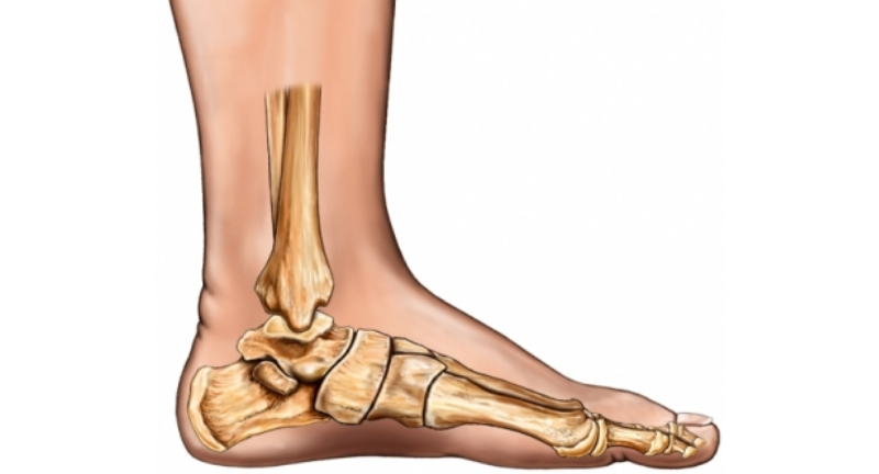
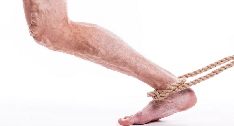
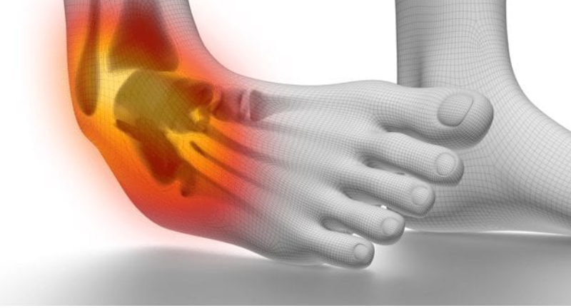
There are several possible causes for the limited range of ankle motion. Often it is due to tightness in the Achilles tendon or calf muscles (the soleus muscle and /or gastrocnemius muscle). In some patients, this tightness is congenital and is sometimes an inherited trait. Other patients acquire the tightness through situations that keep the foot pointing downward for extended periods- such as being in a cast, being on crutches or frequently wearing high heeled shoes. In addition, diabetes can affect the fibers of the Achilles tendon and cause tightness.

Sometimes equinus is related to a bone blocking the ankle motion. For example, a fragment of a broken bone following an ankle injury or bone block can get in the way and restrict motion.

Less often, equinus is caused by spasms in the calf muscle. These spasms may be signs of an underlying neurologic disorder such as cerebral palsy.

Foot Problems related to equinus

Depending on how a patient compensates for the inability to bend properly at the ankle, a variety of foot conditions can develop including:

You’re also more likely to develop an ingrown toenail if you cut your nails at an angle instead of straight across.

Diagnosis

Most patients with equinus are unaware they have this condition when they first visit the doctor. Instead they come to the doctor seeking relief for foot problems associated with the compensation for the short and tight Achilles tendon.

To diagnose equinus, the foot and ankle surgeon will evaluate the ankle’s range of motion when the knee is flexed (bent) as well as extended (straightened). This enables the surgeon to identify whether the tendon or muscle is tight and to assess whether bone is interfering with ankle motion. X-rays may also be ordered. In some cases the foot and ankle surgeon may refer the patient for neurologic evaluation.

Treatment

Non-surgical options

Treatment includes strategies aimed at relieving the symptoms and conditions associated with equinus. In addition, the patient is treated for the equinus itself through one or more of the following options:

Surgical Options

In some cases, surgery may be needed to correct the cause of the short tight Achilles tendon. The foot and ankle surgeon will determine the type of procedure that is best suited to the individual patient.

Plantar Warts

A wart is a small growth on the skin that develops when the skin is infected by a virus. Warts can develop anywhere on the foot, but typically they appear on the bottom of the foot. Plantar warts most commonly occur in children, adolescents, and the elderly. There are two types of plantar warts. A solitary wart is a single wart. If often increases in size and may eventually multiply forming additional satellite warts.

Signs and Symptoms

The signs and symptoms of a plantar wart may include:

Plantar warts grow deep into the skin. Usually, this growth occurs slowly, the art starts off small and gets large over time.

What causes a plantar wart?

Plantar warts are caused by direct contact with the human papilloma virus (HPV). This is the same virus that causes warts on other areas of the body. Typically the plantar wart virus is acquired in public places where people go barefoot. It can also be acquired at home if other family members have the virus.

Diagnosis and Treatment

To diagnose a plantar wart, the foot and ankle surgeon will examine the patient’s foot and look for signs and symptoms of a wart. Although plantar warts may eventually clear up on their own, most patients desire faster relief. The goal of treatment is to completely remove the wart.

The foot and ankle surgeon may use topical or oral treatments, laser therapy, cryotherapy, or surgery to remove the wart. Regardless of the treatment approaches undertaken, it is important that the patient follow the surgeon’s instructions, including all home care and medication that has been prescribed as well as follow up visits with the surgeon. Warts may return requiring further treatment.

If there is no response to treatment, further diagnostic evaluation may be necessary. In such cases, the surgeon can perform a biopsy to rule out other potential causes for the growth.

Rheumatoid Arthritis

It is a disease in which certain cells of the immune system malfunction and attack healthy joints.

RA causes inflammation in the lining of joints, most often the joints of the hands and feet. The signs of inflammation include pain, swelling, redness, and a feeling of warmth around affected joints. IN some patients, chronic inflammation results in damage to the cartilage and bones in the joint. Serious damage can lead to permanent joint destruction, deformity, and disability.

How does RA affect the foot and ankle?

When joints become inflamed due to RA the synovium thickens and produces an excess of joint fluid. This overabundance of fluid, along with inflammatory chemicals released by the immune system, cause swelling and damage to the joint’s cartilage and bones.

Foot problems caused by RA most commonly occur in the forefoot, although RA can also affect other areas of the foot and ankle. The most common signs and symptoms of RA related foot problems in addition to abnormal appearance of deformities are pain, swelling, joint stiffness and difficulty walking.

Deformities and conditions associated with RA may include:

How is RA diagnosed?

Usually, a patient has already been diagnosed with RA prior to visiting the foot and ankle surgeon. However, occasionally a patient first receives a diagnosis of RA from the foot and ankle surgeon. RA is diagnosed on the basis of a clinical examination as well as blood tests. To further evaluate the patient’s foot and ankle problems the surgeon may order x-rays and or other imaging tests.

Treatment of foot and ankle complications seen with RA

While the treatment of RA focuses on the medication prescribed by a patient’s primary doctor or rheumatologist, the foot and ankle surgeon will develop a treatment plan aimed at relieving the pain of RA related foot problems. The plan may include one or more of the following options:

Rheumatoid arthritis in the foot and ankle can cause considerable pain and deformity, making walking difficult. Through the treatment approaches selected by the foot and ankle surgeon substantial relief can be obtained.

Sesamoid Injuries

It is a bone embedded in a tendon. Sesamoids are found in several joints in the body. In the foot, the sesamoids are two pea-shaped bones located in the ball of the foot, beneath the big toe joint. Acting as a pulley for tendons, the sesamoids help the big toe move normally and provide leverage when the big toe “pushes off” during walking and running. The sesamoids also serve as a weight-bearing surface for the first metatarsal bone (the long bone connected to the big toe). Absorbing the weight placed on the ball of the foot when walking, running and jumping.

Types of Sesamoid Injuries in the foot

There are three types of sesamoid injuries in the foot:

Diagnosis

In diagnosing a sesamoid injury, the foot and ankle surgeon will press on the big toe, move it up and down and may assess the patient’s walking and evaluate the wear pattern on the patient’s shoes. X-rays are ordered and in some cases, additional imaging studies such as a bone scan or MRI may also be needed.

Treatment, Non-surgical approaches

Non-surgical treatment for sesamoid injuries of the foot may include one or more of the following options, depending on the type of injury and degree of severity.

When is surgery needed?

Surgery is generally reserved for the most severe sesamoid injuries which fail to respond to non-surgical treatment over a long period of time. In these cases, the foot and ankle surgeon will determine the type of procedure that is best suited to the individual patient.

Peripheral Arterial

Disease (P.A.D)

Commonly referred to as poor circulation PAD is the restriction of blood flow in the arteries of the leg. When arteries become narrowed by plaque (the accumulation of cholesterol and other materials on the walls of the arteries), the oxygen-rich blood flowing through the arteries cannot reach the legs and feet.

Signs & Symptoms

Most people have no symptoms during the early stages of P.A.D. Often, by the time symptoms are noticed the arteries are already significantly blocked. Common Symptoms of P.A.D. include:

If any of these symptoms are present it is important to discuss them with a foot and ankle surgeon. Left untreated P.A.D. can lead to debilitating and limb-threatening consequences.

Risk factors of P.A.D.

Because only half of those with P.A.D. actually experience symptoms, it is important that people with known risk factors be screened or tested for P.A.D. The risk factors include

Diagnosis of P.A.D.

To diagnose P.A.D. the foot and ankle surgeon obtains a comprehensive medical history of the patient. The surgeon performs a lower extremity physical examination that includes evaluation of pulses, skin condition, and foot deformities to determine the patient’s risk of P.A.D. If risk factors are present, the foot and ankle surgeon may order further tests.

Several non-invasive tests are available to assess P.A.D. The ankle-brachial index (ABI) is a simple test in which blood pressure is measured and compared at the arm and ankle levels. An abnormal ABI is a reliable indicator of underlying P.A.D. and may prompt the foot and ankle surgeon to refer the patient to a vascular specialist for additional testing and treatment as necessary.

General Treatment of P.A.D.

Treatment for P.A.D. involves lifestyle changes, medication and in some cases surgery.

P.A.D. and foot problems

Simple foot deformities (hammertoes, bunions, boney prominences) or dermatologic conditions such as ingrown or thickened fungal nails often become more serious concerns when P.A.D is present. Because the legs and feet of someone with P.A.D. do not have normal blood flow and because blood is necessary for healing, seeming small problems such as cuts, blisters or sores can result in serious complications.

Having both diabetes and P.A.D. further increases the potential for foot complications. People with diabetes often have neuropathy (nerve damage that can cause numbness in the feet) so they don’t feel pain when foot problems occur. When neuropathy occurs in people with P.A.D. ulcers can develop over foot deformities and may never heal. For this reason, P.A.D. and diabetes are common causes of foot or leg amputations in the United States.

Once detected P.A.D. may be corrected or least improved. The foot and ankle surgeon can then correct the underlying foot deformity to prevent future problems should the circulation become seriously restricted again.

Avoiding P.A.D. complications

Getting regular foot exams as well as seeking immediate help when you notice changes in the feet-can keep small problems from worsening. P.A.D. requires ongoing attention. To avoid complications, people with this disease should follow these precautions:

Bunions

Even though bunions are a common foot deformity, there are misconceptions about them. Many people may unnecessarily suffer the pain of bunions for years before seeking treatment.

Bunions are often described as a bump on the side of the big toe. But a bunion is more than that. The visible bump reflects changes in the bony framework of the front part of the foot. With a bunion, the big toe leans toward the second toe, rather than pointing straight ahead. This throws the bones out of alignment production the bunion’s bump. Bunions are a progressive disorder. They begin with a leaning of the big toe, gradually changing the ankle of the bones over the years and slowly producing the characteristic bump, which continues to become increasingly prominent. Usually, the symptoms of bunions appear at later stages, although some people never have symptoms.

What causes a bunion?

Bunions are most often caused by an inherited faulty mechanical structure of the foot. It is not the bunion itself that is inherited, but certain foot types that make a person prone to developing a bunion. Although wearing shoes that crowd the toes won’t cause bunions in the first place, it sometimes makes the deformity get progressively worse at a faster rate and will cause more pain around the bunion sight.

Symptoms

Symptoms which occur at the site of the bunion, may include:

Other conditions that may appear with bunions include calluses on the big toe, sores between the toes, ingrown toenails, neuromas (benign swelling of the nerve) between the toes, and restricted motion of the toe.

Diagnosis

Bunions are readily apparent; you can see the prominence at the base of the big toe or the side of the foot. However, to fully evaluate your condition, the podiatric foot and ankle surgeon may take x-ray to determine the degree of the deformity and assess the changes that have occurred, not only around the effected joint but your overall foot structure.

Because bunions are progressive, they don’t go away, and will usually get worse over time. But not all cases are alike, some bunions progress more rapidly than others. Once your podiatric surgeon has evaluated your particular case, a treatment plan can be developed that is suited to your needs.

Treatment

Sometimes observation of the bunion is all that’s needed. Periodic office evaluation and x-ray examination can determine if your bunion deformity is advancing, thereby reducing your chance of irreversible damage to the joint. In many other cases, however. Some type of treatment is needed.

Early treatments are aimed at easing the pain of bunions, but thy will not reverse the deformity itself nor stop the progression.

These options include:

When is surgery needed?

When the pain of a bunion interferes with daily activities or daily shoe wear, it’s time to discuss surgical options with your podiatric surgeon. Together you can decide if surgery is best for you.

Recent advances in surgical techniques have led to a very high success rate in treating bunions.

A variety of surgical procedures are performed to treat bunions. The procedures are designed to remove the bump of bone, correct the changes in the bony structure of the foot as well as correct soft tissue changes that may also have occurred. The goal of these corrections is elimination of pain and provide stability to the foot.

In selecting the procedure or combination of procedures for your particular case, the podiatric surgeon will take into consideration the extent of your deformity based on the x-ray findings, your age, your activity level, overall structure of the foot, as well as other factors. The length of the recovery period will vary, depending on the procedure or procedures performed.

Charcot Foot

Charcot foot is a sudden softening of the bones in the foot that can occur in people who have significant nerve damage (neuropathy). The bones are weakened enough to fracture, and with continued walking the foot eventually changes shape. As the disorder progresses, the arch collapses and the foot takes on a convex shape, giving it a rocker-bottom appearance, making it very difficult to walk. Charcot foot is a very serious condition that can lead to severe deformity, disability, and even amputation. Because of its seriousness, it is important that patients with diabetes a disease often associated with neuropathy take preventive measures and seek immediate care if signs or symptoms occur.

Symptoms

The symptoms of Charcot foot can appear after a sudden trauma or even a minor repetitive trauma (such as a long walk). The symptoms of Charcot foot are like those of an infection. Although Charcot foot and infection are different conditions, both are serious problems requiring medical treatment.

Charcot foot symptoms may include:

What Causes Charcot foot?

Charcot foot develops as a result of neuropathy, which decreases sensation and the ability to feel temperature, pain or trauma. When neuropathy is severe, there is a total lack of feeling in the feet. Because of neuropathy, the pain of an injury goes unnoticed and the patient continues to walk making the injury worse.

People with neuropathy are at risk of developing Charcot foot. In addition, neuropathic patients with a tight Achilles tendon have been shown to have a tendency to develop Charcot foot.

Diagnosis

Early diagnosis of a Charcot foot is extremely important for successful treatment. To arrive at a diagnosis, the surgeon will examine the foot and ankle an ask about events that may have occurred prior to the symptoms.

X-rays are also essential for diagnosis. In some cases, other imaging studies and lab test may be ordered. Once treatment begins, x-rays are taken periodically to aid in evaluating the status of the condition.

Treatment

It is extremely important to follow the surgeon’s treatment plan for Charcot foot. Failure to do so can lead to the loss of a toe, foot, leg or life.

Treatment for Charcot foot consists of:

Preventive Care

The patient can play a vital role in preventing Charcot foot and its complications by the following measures.

Flat Foot

It is a complex disorder, with diverse symptoms and varying degrees of deformity and disability. There are several types of flatfoot, all of which have one characteristic in common-partial or total collapse (loss) of the arch. Other characteristics shared by most types of flatfoot include toe drift, where the toes and front part of the foot point outward, the heel tilts toward the outside and the ankle appears to turn in, a short Achilles tendon, which causes the heel to lift off the ground earlier when walking and may act as a deforming force, bunions and hammertoes may occur in some people with flatfeet, health problems such as rheumatoid arthritis or diabetes sometimes increase the risk of developing flatfoot. In addition, adults who are overweight frequently have flatfoot.

Flexible Flatfoot

Flexible flatfoot is one of the most common types of flatfoot. It typically begins in childhood or adolescence and continues into adulthood. It usually occurs in both feet and generally progresses in severity through the adult years. As the deformity worsens, the soft tissues of the arch may stretch or tear and can become inflamed. This usually involves the posterior tibial tendon and the deltoid ligament complex.

The term flexible means that while the foot is flat when standing (weight-bearing), the arch returns when not standing. In the early stages of flexible flatfoot, arthritis is not restricting motion of the arch and foot, but in the later stages, arthritis may develop to such a point that the arch and foot become stiff.

Symptoms

Diagnosis

In diagnosing flatfoot, the podiatric foot and ankle surgeon examines the foot and observes how it looks when you stand and sit. X-rays are taken to determine the severity of the disorder. If you are diagnosed with flexible flatfoot but you do not have any symptoms, your podiatric surgeon will explain what you might expect in the future.

Treatment

If you experience symptoms with flexible flatfoot the podiatric surgeon may recommend various treatment options, including:

Flatfoot Surgery

A variety of surgical techniques is available to correct flexible flatfoot. Your case may require one procedure or a combination of procedures. All of these surgical techniques are aimed at relieving the symptoms and improving foot function. Among these procedures are tendon transfers or tendon lengthening procedures. Realignment of one or more bones, joint fusions or insertion of implant devices.

In selecting the procedure or combination of procedures for your particular case, the podiatric surgeon will take into consideration the extent of your deformity base on the x-ray findings, your age, your activity level, and other factors. The length of the recovery period will vary, depending on the procedure or procedures performed.